Korva is a scroll of the internet’s best ideas.
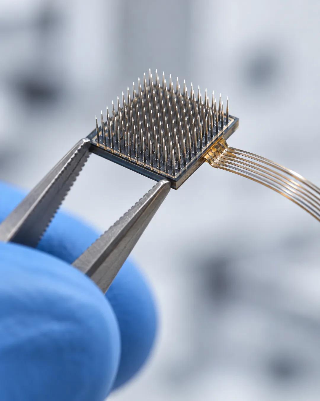
Reading a brain means breaching the skull

How Neurons Signal
Neurons communicate through action potentials, electrical signals. Textbooks teach that these are always the same size and shape for a given neuron, which turns out not to be true. A neuron either fires or it does not, but graded potentials matter: once a threshold is reached the neuron fires and communicates with the next one, depending on how many other inputs there are and what they are doing.
Ways to Record
Recording from neurons can mean putting an electrode close to one, laying a bed of nails nearby to pick up lots of extracellular signals, or impaling a neuron to record from inside it. All of that breaches the skull and sticks wires into the brain. At the other end sit fMRI and EEG, the heat maps of the brain, which only measure signals on the surface at the cortex and cannot reach the deeper layers.
Noninvasive Methods
Ultrasound and transcranial magnetic stimulation, which is used for the treatment of depression, are the exciting noninvasive methods. For stimulation, the spatial control of transcranial magnetic stimulation is not great.
Why can't fMRI see individual neurons?
Functional MRI does not record electrical activity. It tracks changes in blood oxygen, which follow neural activity by a few seconds. One imaging voxel, a cube typically a few millimeters on a side, contains hundreds of thousands to millions of neurons. The signal therefore shows where activity increased, not which individual cells fired.
How does magnetic stimulation excite neurons?
Transcranial magnetic stimulation passes a brief current through a coil held against the scalp, creating a rapidly changing magnetic field. By Faraday's law of induction, that field induces a small electric current in the cortex underneath. The current can make neurons there fire. The US Food and Drug Administration cleared the first TMS device for depression in 2008.
Why does EEG only see the brain's surface?
EEG electrodes sit on the scalp, and the skull and other tissue blur and weaken the electrical fields passing through. What they pick up best is the synchronized activity of thousands of cortical neurons aligned in the same direction. Signals from deep structures are too small and diffuse to separate. The spatial resolution of scalp EEG is on the order of centimeters, while its timing resolution is in milliseconds.
More from Invest Like the Best with Patrick O'Shaughnessy