A Wet DNA Photo Helped Reveal a Double Helix
Wet DNA gave sharp X-ray spots showing two helices, and measurements from them pointed to paired rungs inside.
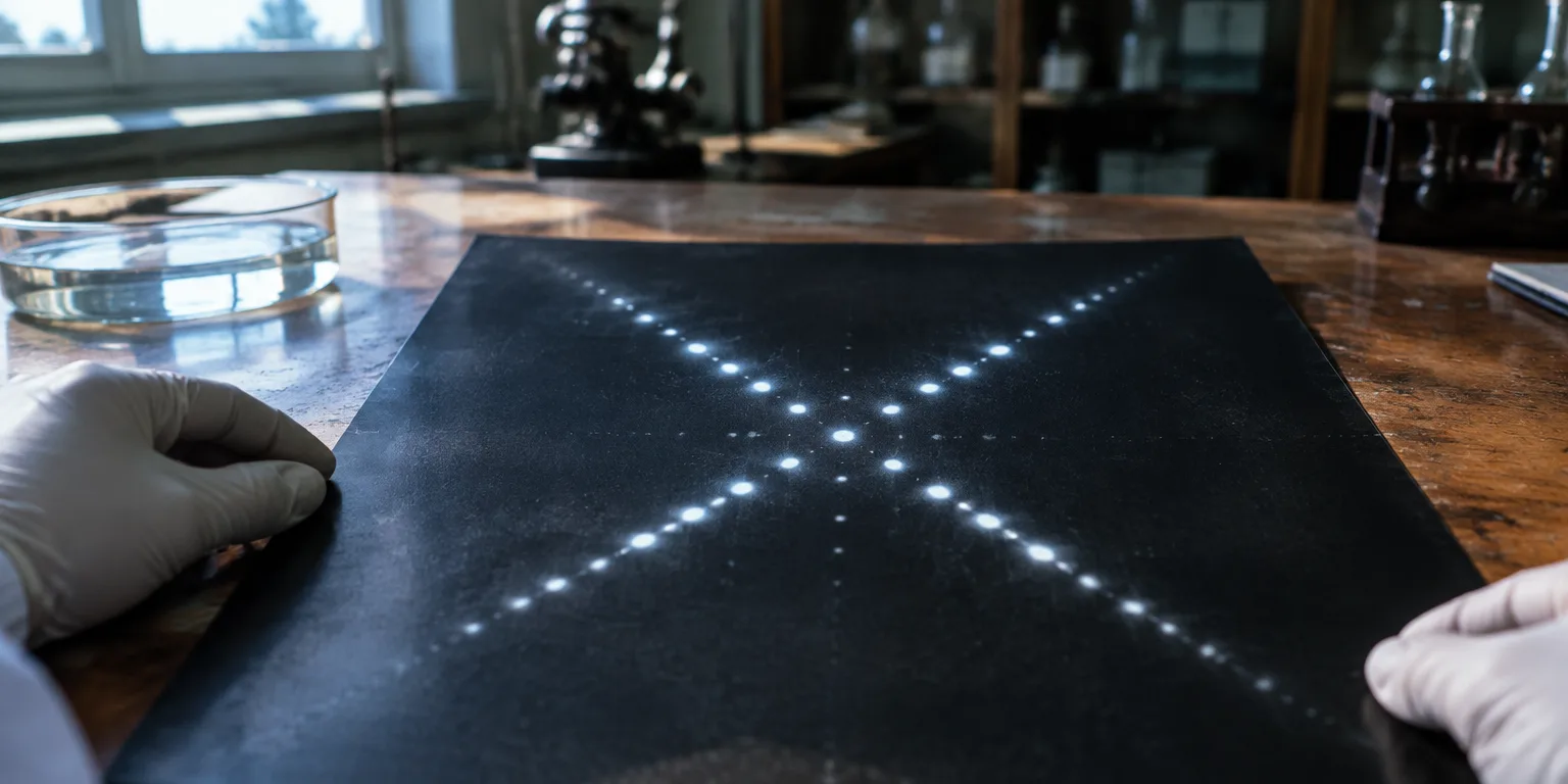
The secret of how genes copy themselves came into focus when scientists added water to DNA. The damp sample gave a sharp X-ray image that revealed a double spiral.
In the 1930s, the favored guess for the material of genes was protein. That view shifted after 1944 lab work in New York showed that DNA, not protein, could transform bacteria.
DNA proved to be a hefty structure built from sugar, phosphate, and four smaller chemicals called bases, adenine, cytosine, guanine, and thymine. Biochemist Erwin Chargaff then noticed that the amount of adenine usually matched thymine, and cytosine matched guanine, as if each had a partner.
Seeing the partners required a clearer picture. At King's College London, physicist Maurice Wilkins working with young researcher Raymond Gosling learned to control moisture in DNA, creating a perfectly orderly sample that gave far sharper X-ray patterns than earlier blurry images.
In X-ray work, beams are aimed at the sample and bend as they pass through, and the positions and strengths of the scattered rays are recorded on film. With the fully hydrated form of DNA, known as the B form, that wet-DNA image, nicknamed Photo 51, revealed a pair of intertwined strands and made precise measurement of the spots possible.
Those measurements pointed researchers at Cambridge, James Watson and Francis Crick, to the ladder. The sugar phosphate chains formed the outside rails and ran in opposite directions, while the paired bases met them at right angles to form the rungs.
Watson and Crick drew the copying idea from the pairing rules. If the two rails pulled apart, each exposed row of bases could serve as a guide for building a new matching chain, handing down biological directions to offspring.
Questions people ask
How can an X-ray picture show the shape of a molecule?
X-rays are aimed at an orderly sample and bend as they pass through, and the scattered rays are recorded as spots on film. The positions and brightness of the spots reveal distances and angles inside the molecule. For DNA, a wetter and more orderly sample gave sharper spots that could be measured precisely.
Why did wet DNA give a clearer picture than dry DNA?
Water helps long DNA molecules line up neatly and take on a regular spiral shape. That order turns a fuzzy blur into a crisp pattern with measurable spots. Researchers could then calculate the width of the helix and the spacing of the rungs.
Which bases pair together, and why does it matter?
Adenine pairs with thymine, and cytosine pairs with guanine. Each pair has about the same width, so the ladder keeps a uniform shape. The pairing rule also lets each strand act as a template for building a new partner strand.