New Study Suggests Rosalind Franklin Saw DNA's Spiral First
She started the sharp Photo 51 the same day she saw the rough Photo 49, a sign she knew the spiral clue was important.
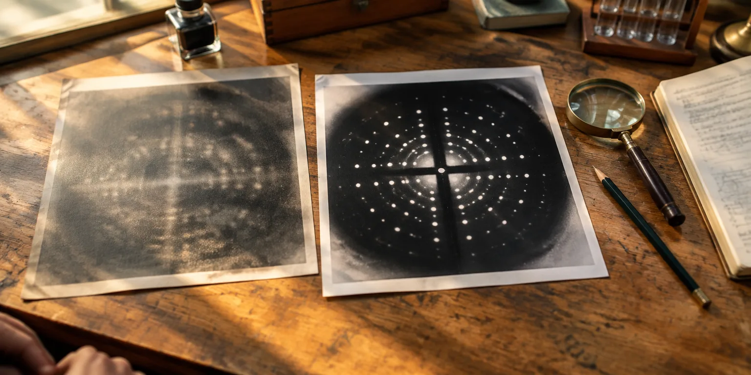
A new study of her notes and X-ray images argues Rosalind Franklin likely saw DNA's spiral shape before Watson and Crick published the double helix. She reshot a rough image to make the sharp picture known as Photo 51.
Photo 51, taken in May 1952, was a fresh exposure of the DNA sample she had already photographed as Photo 49, started the same day she saw the old one. Study coauthor Alistair Sponsel, a historian of science at the Science History Institute in Philadelphia, says she wanted a more refined version.
Her notebooks support that reading. Notes on Photo 49 mention a possible spiral shape and rich structural detail. The study was published in the Journal of the History of Biology.
Retaking the same sample suggests she knew the spiral clue mattered. James Watson, who later shared the Nobel Prize for the double helix, wrote the black cross at Photo 51's center could only have come from a spiral structure.
Franklin used X-ray imaging. The method maps molecules by scattering X-rays off them. Sponsel reads Photo 49 as the experiment that found promising data, and Photo 51 as the showcase copy made to show those findings with maximum clarity.
Questions people ask
What exactly is an X-ray photo of DNA?
It is an X-ray diffraction picture, made by aiming X-rays at DNA fibers and recording how the rays scatter. The scatter forms a pattern of spots and crosses, and scientists work backward from that pattern to infer the molecule's three-dimensional shape.
Why didn't Franklin share the Nobel Prize?
Franklin died in 1958, four years before Watson, Francis Crick, and Maurice Wilkins shared the 1962 Nobel Prize in Medicine for the double helix. Nobel Prizes are not awarded posthumously, and Watson's later memoir cast her as skilled in technique but slow to interpret her images, a view the new archival work disputes.
What is B-form DNA?
The B-form is the water-rich form of DNA, and it is the form our cells normally use. Franklin also studied the drier A-form because its images looked richer in data, and she planned to characterize A fully before turning back to B.