Rosalind Franklin Kept Solving Structures, From Coal to DNA to Viruses
She left King's College unhappy, built a leading virus lab, and died before the reviews of her Brussels model appeared.
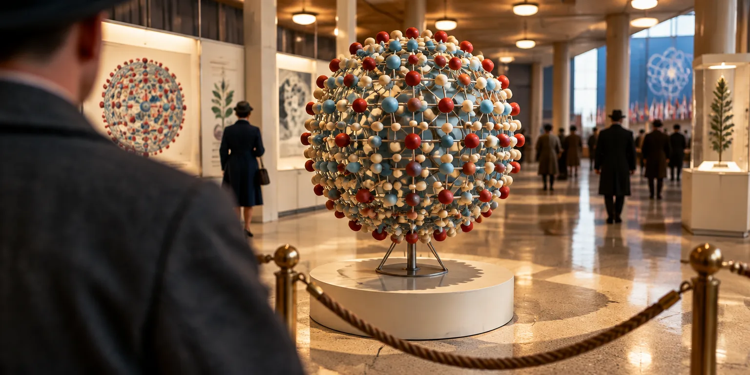
Visitors to the international exposition in Belgium in 1958 saw a large demonstration model of a plant virus.
Its builder, chemist Rosalind Franklin, had ovarian cancer and died in 1958 before the favorable reviews appeared.
Franklin built a short career on using X-rays to uncover how matter is put together, starting with coal, then DNA, and then viruses.
She was born July 25, 1920 in London to a well-known Jewish household. Her parents saw schooling as a path to personal and social welfare, and as a girl she joined her father Ellis in supporting a college that held night lessons for workers.
She finished an elite girls school ahead of schedule, then studied physical chemistry at Newnham College, a women's college of Cambridge University. During World War II she wanted to aid the war effort without leaving research, and welcomed a post at the British Coal Utilization Research Association.
There she investigated the qualities of coal and graphite. That research supplied her 1945 doctorate and material for her earliest papers.
After the war she moved to Paris for postdoctoral work at the Laboratoire Centrale des Services Chimiques de l'Etat. She received thorough instruction in X-ray crystallography there and soon earned a reputation as a specialist.
The method directs X-rays at crystals and captures how the rays scatter on film, revealing atomic positions. Biophysicist John T. Randall, who led the biophysics unit at King's College London, sought that expertise. His unit counted 8 women among 31 researchers, an unusually high share then, and he offered Franklin a post in 1951.
Colleagues Maurice Wilkins and his pupil Raymond Gosling were probing DNA with a remarkably clean sample from chemist Rudolf Signer, drawn from calf thymus, that could be pulled into slender fibers yielding sharp patterns. Her arrival blurred who oversaw which subjects and methods, stirring friction in the laboratory.
Then came a strikingly sharp picture of B-type DNA, the form of DNA that is fully wet with water inside it, later called Photo 51. It revealed a double spiral and its spots were clear enough for detailed measurement of scattered strength.
From the pattern, James Watson and Francis Crick, two researchers at Cambridge University who built a DNA model, inferred outer sugar-phosphate backbones twisting opposite ways, with paired bases crossing them at right angles at set gaps. The shape suggested the split strands could act as a guide for making new proteins.
Unhappy at King's, Franklin departed shortly after making the picture for the laboratory of J. D. Bernal, a crystallographer, a scientist who studies crystal structures by X-ray, who ran a lab at Birkbeck College in London. There she turned tools honed on DNA to a plant virus and to the polio virus, advancing knowledge of their makeup.
Her laboratory thrived because she won access to choice specimens, instruments, and colleagues' results, and she kept cordial ties with Watson and Crick. Public notice arrived when Lawrence Bragg, co-developer of X-ray crystallography with his father William, asked her to build the big demonstration model of a plant virus for Brussels.
Ovarian cancer ended her life in 1958. Watson and Crick had unveiled the double helix in a brief 1953 Nature report, and nine years later shared the Nobel Prize for Physiology or Medicine with Wilkins. Custom then barred posthumous awards, so she was omitted from the honor.
Questions people ask
How does X-ray diffraction reveal a structure?
X-rays are aimed at a crystal and bend as they pass through it. The positions and brightness of the diffracted rays are recorded on film. From those spots, scientists calculate where atoms sit and what repeating shapes they form.
What was Photo 51?
Photo 51 was an X-ray image of the fully hydrated, or B form, of DNA. It clearly showed two helices and its intensities could be measured with great precision. Watson saw the image after Wilkins showed it to him, and those measurements helped fix the spacing and direction of the two chains.
What are tobacco mosaic virus and poliovirus?
Tobacco mosaic virus infects tobacco and related plants, mottling the leaves, and it became a laboratory workhorse because it is abundant and sturdy. Poliovirus infects people and can cause polio. Franklin's team studied how their protein and nucleic acid parts were arranged.
Why was Franklin not included in the 1962 Nobel Prize?
Nobel Prizes were traditionally not awarded posthumously, so a person who had died could not be chosen. Franklin died in 1958 and the DNA prize was awarded in 1962 to Watson, Crick, and Wilkins. Historians still debate whether she would have shared it had she lived.